What is a cerebral angiogram?
A cerebral angiogram is an imaging technique used to see how blood flows in the arteries and veins of the brain in real time.
During an angiogram, a catheter is placed in the femoral artery in the groin and advanced to the main artery in the neck. Dye is injected into the artery, which is then visualized up by a fluoroscope
Besides the arteries, an angiogram is able to show all of the blood vessels in your brain as the contrast (dye) flows from your arteries to your veins.
Since an angiogram is a video and not a single picture, blood flow can be observed in different phases. The two main phases are the arterial phase and the venous phase. Blood flows into the brain during the arterial phase and out of the brain during the venous phase.
We have 3 convenient locations throughout Southeastern Wisconsin.
What should I expect during a cerebral angiogram?
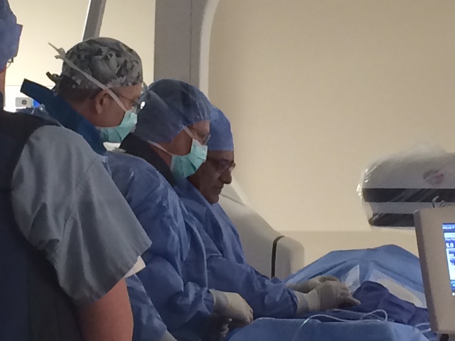
Dr. Ahuja performs the angiograms himself. It is an outpatient procedure, so patients will likely go home later the same day. The entire procedure takes about a half hour.
While patients are not put to sleep, they are given a sedative to help them stay relaxed. Dr. Ahuja will also numb the area so patients don’t feel pain.
At the start of the procedure Dr. Ahuja places an IV line near the groin, in the femoral artery. Once Dr. Ahuja has access to the artery he threads a thin, flexible plastic tube called a catheter (only about 2 millimeters in diameter) into the femoral artery. Using the special x-ray machine as guidance, he thread the catheter up into your carotid artery, in the neck.
Once in the artery, Dr. Ahuja injects a small amount of contrast dye into the bloodstream, which travels through the brain and shows how the blood flows. He may do this several times to get different images.
When the dye is injected, patients are asked to hold their breath and remain still for a short period of time to make sure the picture is clear. Patients may feel slightly warm or flushed when the dye is injected, which is perfectly normal.
If patients have an iodine or seafood allergy, they should advise Dr. Ahuja because the contrast material contains iodine. Medication can be given to compensate for this prior to the procedure.
If at any time during or after the procedure a patient experiences numbness, weakness, dizziness, chest pain, slurred speech, blurry vision, or trouble breathing, they should let the staff at Dr. Ahuja’s office know immediately.
This information is presented to our patients many times before the procedure, and we want you to be comfortable with every aspect. You will have a chance to ask any questions that are on your mind.
What is a biplane angiography system?
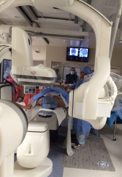
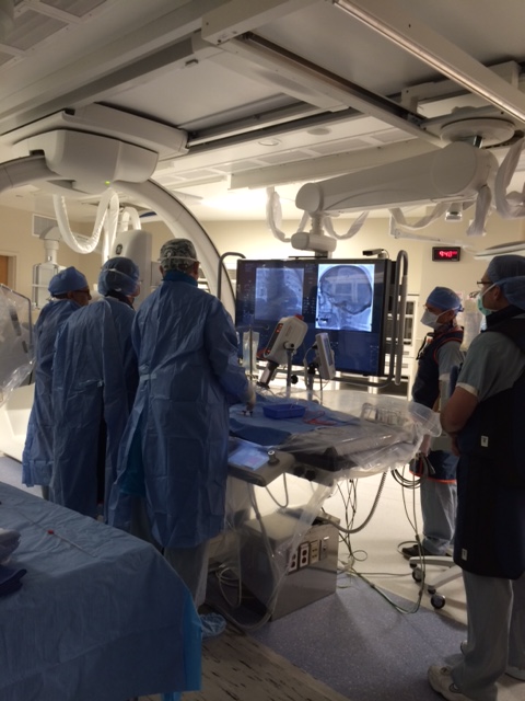
Dr. Ahuja recently inaugurated a new state-of-the-art biplane angiography system at St. Catherine’s Hospital in Pleasant Prairie, Wisconsin (near Kenosha).
This new system allows for the diagnosis and treatment of diseases such as brain aneurysms, stroke, tumors, and other neurological conditions.
Dr. Ahuja has been performing these procedures for over 20 years, but this latest technological improvement allows him to provide cutting edge care for patients.


